
What Is Sialography?
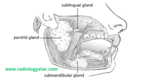
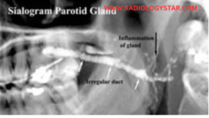
Sialography is the radiological examination to help the study of salivary glands ( parotid gland, submandibular, sublingual gland) by the injection of contrast media through the orifice or opening of duct.

Indication Of Sialography
— Pain
— Swelling
— Calculus.
— Mass lesions.
— Obstructive lesions.
— Penetrating trauma.
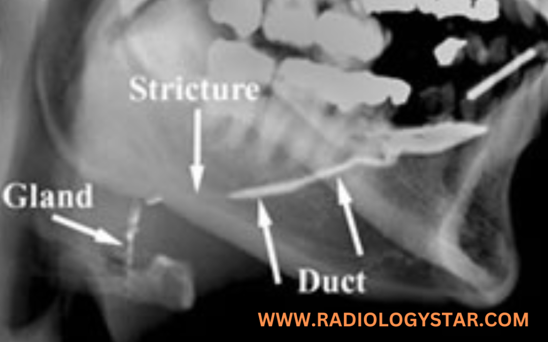
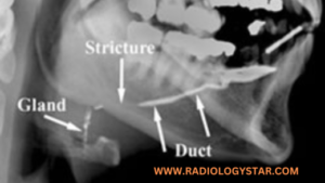
— Stricture.
— Fistula.
— Sialectasis
— Acute Peritonitis.
— Sialoria.
Contraindications.
— Acute and chronic inflammation.
— Sensitivity to contrast media.
Contrast media.
— HOCM / LOCM ( Lipiodol 240-300 mg)
— Dose:- 2-3 ml.
Equipment.
— Skull unit.
— Fluoroscopy unit.
— Lemon.
— Anesthesia.
–Cannula.
— Syringe 2cc.
Patient preparation.
— The must be removed all radio- opaque equipment from the examination area. Like false teeth, ornaments,
— Must be fill the consent from.
Preliminary Film.
A) For Parotid gland.
— AP view of face with 5° away from examination area
— Lateral with centering at the angle of mandible.
— Lateral oblique 20° cephalic and centring at the angle of mandible.
B) Submandibular gland
— Infero – superior views using occlusal film.
— Lateral with floor of mouth depressed with a wooden spatula.
— Lateral oblique centering 1 cm anterior to the angle of mandible.
Procedure / Technique.
— The patient lies supine position on the fluoroscopy table.
— To ask the patient for open the mouth.
— The anesthesia spray apply on the salivary duct for the patient make relaxation.
— The orifice of the parotid duct is present to the crown of the second upper molar.
— The orifice of the submandibular duct present in the base of frenulum of the tongue.
— If the orifice or opening of ducts are not seen then some amount of lemon ( citric acid) is placed under the mouth for secretion of saliva and orifice visible.
— Then the duct is dilated to the help of dilator and the cannula or catheter is introducing in to the duct.
— The 1-2 ml of Contrast is injected in to duct.
— If the any types of pain is occurs then the contrast injection is stop.
— Then taken a spot film immediately and after taking a film the cannula or catheter are removed.
Filming.
A) For Parotid gland.
— AP view of face with 5° away from examination area
— Lateral with centering at the angle of mandible.
— Lateral oblique 20° cephalic and centring at the angle of mandible.
B) Submandibular gland
— Infero – superior views using occlusal film.
— Lateral with floor of mouth depressed with a wooden spatula.
— Lateral oblique centring 1 cm anterior to the angle of mandible.
C) Post – Secretory film.
— After 5 min injected of contrast media the post secretory film taken.
Complications.
— Pain
— Damage to the duct orifice.
— Rupture of the ducts.
— Infection.
Aftercare.
— The patient keep in the observation for 2-3 hrs for any types of complications.
FAQs.
Q. What is sialography?
Sialography is a medical imaging technique used to visualize the salivary glands and ducts using X-rays and a contrast agent.
Q. Why is sialography performed?
Sialography is performed to diagnose salivary gland and duct abnormalities, such as stones, tumors, or strictures.
Q. How is sialography performed?
A contrast agent is injected into the salivary ducts, and X-ray images are taken as the contrast agent flows through the glands and ducts.
Q. What is the contrast agent used in sialography?
Commonly, a water-soluble iodinated contrast agent is used.
Q. Is sialography painful?
The injection may cause discomfort, but the procedure itself is not usually painful.
Q. Are there any risks associated with sialography?
There is a slight risk of infection, allergic reactions to the contrast agent, and radiation exposure from X-rays.
Q. How long does a sialography procedure take?
The procedure usually takes about 30 to 60 minutes.
Q. Can sialography detect salivary gland tumors?
Yes, sialography can help identify tumors and their location within the salivary glands.
Q. What is sialolithiasis?
Sialolithiasis is the formation of salivary gland stones that can block ducts.
Q. Can sialography diagnose sialolithiasis?
Yes, sialography can reveal the presence and location of salivary gland stones.
Q. Are there alternatives to sialography?
Yes, ultrasound, CT scans, and MRI are alternative imaging methods for salivary gland evaluation.
Q. Is sialography suitable for all patients?
Sialography may not be suitable for pregnant individuals and those with severe allergies to contrast agents.
Q. Is sedation necessary for sialography?
Sedation is not typically required for sialography.
Q. How should I prepare for sialography?
Follow any fasting or fluid intake instructions provided by your doctor.
Q. Can sialography diagnose autoimmune disorders affecting salivary glands?
Sialography may not be the best method for diagnosing autoimmune disorders. Biopsy and blood tests are often used for this purpose.
Q. How do I find a qualified medical professional to perform sialography?
Consult your primary care physician or an oral and maxillofacial radiologist.
Q. Can children undergo sialography?
Yes, children can undergo sialography if necessary, under proper medical guidance.
Q. Is sialography available in all medical facilities?
Sialography might not be available in all facilities; larger medical centers or institutions are more likely to offer this procedure.
Q. Is sialography covered by medical insurance?
Check with your insurance provider to determine if sialography is covered under your plan.
Q. Can sialography diagnose salivary gland infections?
Sialography might not be the primary choice for diagnosing infections. Other imaging methods and clinical assessments are often used.
Q. Is sialography uncomfortable after the procedure?
Discomfort is typically minimal after the procedure and should subside quickly.
Q. How soon can I resume normal activities after sialography?
You can generally resume normal activities immediately after the procedure.
Q. Are there any long-term effects of sialography?
There are usually no long-term effects associated with sialography.
Q. Can sialography be used to guide surgical procedures?
Yes, sialography findings can help guide surgical interventions if necessary.
Q. Can salivary gland stones pass naturally after sialography?
In some cases, small salivary gland stones may pass naturally after the procedure.
Q. What information can sialography provide that other imaging methods can’t?
Sialography provides detailed images of the ducts’ structure and flow of contrast agent, which can be beneficial for specific diagnostic cases.
Q. Is sialography commonly performed today?
Sialography is less common due to the availability of alternative imaging methods, but it can still be valuable in certain scenarios.
Q. Can sialography detect blocked salivary ducts?
Yes, sialography can detect blockages and structural issues in salivary ducts.
Q What should I expect during the sialography procedure?
You will be positioned for X-rays, the contrast agent will be injected, and X-ray images will be taken as it flows through your salivary glands.
Q. How quickly will I receive the results of my sialography?
Your doctor will review the images and discuss the results with you shortly after the procedure.
FORE MORE SPECIAL PROCEDURE CLICK HERE
BOOK LINK :- Fundamentals of Special Radiographic Procedures