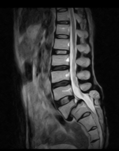
What Is Anterolisthesis?
Anterolisthesis is also called Spondylolisthesis. Anterolisthesis is a medical condition characterized by the forward displacement of one vertebra in relation to the vertebra below it. This displacement typically occurs in the spine, particularly in the lumbar (lower back) region. The term is derived from the Greek words “ante,” meaning “front,” and “listhesis,” meaning “slip.”
Anterolisthesis can result from various causes, including degenerative changes in the spine, trauma, congenital abnormalities, or conditions such as spondylolysis (a defect in the vertebral arch) and spondylolisthesis (a condition where one vertebra slips forward over the one below it). The severity of anterolisthesis is often graded on a scale from 1 to 4, with higher grades indicating more significant displacement.
The term “listhesis” means to slip forward. It occurs when the weakened pars interarticularis separates and allows the vertebra to move forward out of position causing pinched nerves and pain.
Classification of Anterolisthesis.
Anterolisthesis is often classified based on the degree of slippage or displacement of the affected vertebrae. The grading system used is usually a numeric scale ranging from 1 to 4, with each grade indicating the severity of the slippage.
1. Grade I: 1 – 25 % forward slip
2. Grade II: 26 – 50 % forward slip
3. Grade III: 51 – 75 % forward slip
4. Grade IV: 76 – 99% forward slip
What are the symptoms?
1. Back pain
2. Stiffness
3. Muscle Spasms
4. Neurological symptoms
5. Change in posture and Gait
6. Leg pain
How Is A Diagnosis Made?
1. Medical History:- The healthcare provider will begin by obtaining a detailed medical history, including information about the patient’s symptoms, the onset of pain, any injuries or trauma, and any relevant family history of spine-related issues.
2. Physical Examination:- A thorough physical examination is conducted to assess the patient’s range of motion, neurological function, and any signs of muscle weakness, sensory changes, or reflex abnormalities.
3. X-rays:- X-rays are commonly used to visualize the spine and can help identify the presence of anterolisthesis, its severity, and the affected vertebrae. X-rays provide valuable information about the alignment and stability of the spine.

4. MRI (Magnetic Resonance Imaging):- MRI scans provide detailed images of soft tissues, nerves, and discs in the spine. This imaging modality can help evaluate any nerve compression, spinal cord abnormalities, or soft tissue damage associated with anterolisthesis.

5. CT (Computed Tomography):- CT scans may be used to provide more detailed images of the bones and can help assess the degree of slippage and any associated bony abnormalities.

6. Additional Tests:- In some cases, additional tests such as electromyography (EMG) or nerve conduction studies may be conducted to assess nerve function and identify any nerve compression.
BOOK LINK :- Radiographic Pathology for Technologists

