
what Is An X-ray?
X-rays are a type of electromagnetic radiation with wavelengths shorter and higher energies. They were discovered by Wilhelm Conrad Roentgen in 8 NOV 1895. X-rays are produced when high-energy electrons collide with a target material, causing the electrons to undergo rapid deceleration. This deceleration results in the emission of X-ray photons.
X-rays have the ability to penetrate various materials, including human tissues, depending on the material’s density and thickness. This property makes X-rays valuable for imaging the internal structures of objects, from the human body to industrial components. The images produced by X-rays are known as X-ray radiographs or X-ray images
In the medical field, X-rays are widely used for diagnostic purposes. They can pass through soft tissues in the body, but denser materials like bones and certain metals absorb them, creating contrast in the resulting image. This makes X-rays particularly useful for detecting fractures, evaluating the health of bones, and diagnosing various medical conditions.
X-rays are also used in fields such as astronomy, materials science, and industrial applications. In astronomy, X-ray telescopes are used to observe high-energy phenomena in space, such as black holes and neutron stars. In materials science, X-ray diffraction and X-ray fluorescence techniques are used to analyze the composition and structure of materials at the atomic level. In industry, X-rays are employed for quality control and non-destructive testing of manufactured products.
Types of x-ray
There are several different types of X-ray exams that are used for different purposes:
A. Plain film X-ray:- This is the most common type of X-ray exam. It is used to visualize bones and the spaces between them, as well as to look for abnormalities such as fractures or bone tumors.
B. Fluoroscopy:- This type of X-ray exam uses a continuous beam of radiation to produce real-time images of the inside of the body. It is often used to visualize the digestive system during an upper gastrointestinal (UGI) or lower gastrointestinal (LGI) series, or to guide medical procedures such as catheterization.
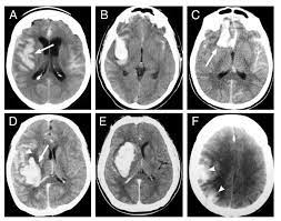
C. Computed tomography (CT) scan:- This is a specialized X-ray exam that uses a series of cross-sectional images to create detailed 3D images of the inside of the body. CT scans are often used to visualize the brain, spine, chest, and abdomen.
D. Mammography:- This is a specialized X-ray exam used to visualize the breasts. It is used to detect breast cancer and other abnormalities in the breast tissue.

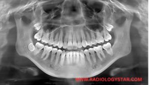
E. Dental X-ray:- This type of X-ray exam is used to visualize the teeth and surrounding structures in the mouth. It is often used to diagnose tooth decay, abscesses, and other dental problems.
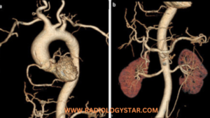
F. Angiography:- This is a specialized X-ray exam used to visualize the blood vessels in the body. It is often used to diagnose problems with the arteries and veins, such as blockages or aneurysms.
Properties of x-ray
Properties of x-ray are given below
1) X-ray are invisible.
2) X-ray travel in straight line.
3) X-ray are not affected by electrical or magnetic fields.
4) X-ray have great penetration power.
5) Being electromagnetic wave, X-ray are electrically in neutral
6) X-ray affect photographic emulsion.
7) X-ray can liberate photoelectrons.
8) X-ray are partially scattered by matter
9) X-ray are able to ionize matter.
10) X-ray are cause fluorescence of certain crystal.
11) Their wave length ranges from 0.1-0.5 A and energy levels at 25Kev to 125 Kev.
12) X-ray travel in a straight line like 3 x 10⁻⁸ m/s.
13) X-ray cause biological change in living organism.
14) X-ray can produce secondary and scattered radiation.
15) It cannot be focused by a lens.
16) it effect photography film and can produce latent image.
Uses Of X-ray In Radiology
Here are some of the main uses of X-ray in radiology:
A. Bone Imaging:- X-rays are excellent for visualizing bones and skeletal structures. They are used to detect fractures, dislocations, and bone diseases like osteoporosis. X-rays can also help orthopedic surgeons during procedures such as setting broken bones.
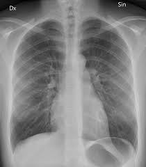
B. Chest Radiography:- Chest X-rays are commonly used to examine the lungs and heart. They can reveal conditions like pneumonia, lung tumors, congestive heart failure, and other abnormalities.
C. Dental X-rays:- Dentists use X-rays to diagnose dental issues such as cavities, gum disease, and impacted teeth. Dental X-rays can also help plan treatments like orthodontic work and dental implants.
D. Mammography:- Mammograms are specialized X-ray exams for breast cancer screening and diagnosis. They can detect breast tumors at an early stage, when they are too small to be felt.
E. Fluoroscopy:- Fluoroscopy involves continuous X-ray imaging in real-time. It’s used for procedures like barium swallow tests (to examine the digestive system) and angiography (to visualize blood vessels).
F. Gastrointestinal Imaging:- X-rays with contrast agents are used to visualize the digestive system. Barium contrast studies can show abnormalities in the esophagus, stomach, and intestines.
G. Urinary Tract Imaging:- X-rays with contrast agents can help visualize the urinary system, identifying kidney stones, tumors, and other conditions.
H. Interventional Radiology:- X-ray guidance is used during minimally invasive procedures to guide catheters, needles, and other instruments to specific areas within the body. This includes procedures like angioplasty, stent placement, and biopsies.
I. Orthopedic Imaging:- X-rays help orthopedic specialists diagnose and plan treatments for various musculoskeletal conditions, including joint problems, fractures, and deformities.
J. Trauma Assessment:- X-rays are used to assess injuries after accidents or trauma, helping doctors determine the extent of damage and the best course of action.
K. Pediatric Imaging:- X-rays are used for diagnosing pediatric conditions, often with reduced radiation doses to minimize risks to young patients.
L. Lung Imaging:- X-rays are used to diagnose lung diseases like tuberculosis, lung cancer, and chronic obstructive pulmonary disease (COPD).
FAQs.
Q. What are X-rays?
X-rays are a type of electromagnetic radiation with shorter wavelengths and higher energies than visible light.
Q. How were X-rays discovered?
X-rays were discovered by Wilhelm Conrad Roentgen in 1895 while experimenting with cathode rays.
Q. How are X-rays produced?
X-rays are produced when high-energy electrons collide with a target material, causing the electrons to decelerate and emit X-ray photons.
Q. What is the main application of X-rays in medicine?
X-rays are widely used in medicine for diagnostic imaging to visualize bones, organs, and tissues.
Q. How do X-rays interact with matter?
X-rays can be absorbed, scattered, or transmitted when they pass through matter. Dense materials like bones absorb more X-rays.
Q. What is the difference between hard and soft X-rays?
Hard X-rays have higher energies and shorter wavelengths, while soft X-rays have lower energies and longer wavelengths.
Q. Are X-rays harmful?
Excessive exposure to X-rays can be harmful as they are ionizing radiation, but proper safety measures are taken in medical and industrial settings.
Q. What is a radiograph?
A radiograph is an image produced by X-rays passing through an object and being captured on a detector, creating a shadow-like image.
Q. What is a contrast agent in X-ray imaging?
A contrast agent is a substance introduced into the body to enhance the visibility of specific structures during X-ray imaging.
Q. How is a mammogram different from a regular X-ray?
A mammogram is a specialized X-ray used for breast imaging, typically with lower radiation doses.
Q. What is the purpose of fluoroscopy?
Fluoroscopy is real-time X-ray imaging used during medical procedures to visualize moving structures, like the digestive tract or blood vessels.
Q. What is an X-ray tube?
An X-ray tube is a device that generates X-rays by accelerating electrons and directing them onto a target material.
Q. How do X-rays help in diagnosing fractures?
X-rays can visualize fractures by showing breaks or discontinuities in bone structures.
Q. What is computed tomography (CT) scanning?
CT scanning is a technique that uses X-rays to create detailed cross-sectional images of the body, providing more information than traditional X-rays.
Q. How is radiation exposure minimized during X-ray procedures?
Radiation exposure is minimized by using the lowest possible dose while maintaining image quality and following safety guidelines.
Q. Can pregnant women have X-rays?
Pregnant women should avoid unnecessary X-rays due to the potential risk to the developing fetus, but in emergencies, proper shielding can be used.
Q. What is the difference between X-ray and MRI?
X-rays use ionizing radiation and are best for visualizing bones, while MRI uses strong magnetic fields and radio waves to create detailed images of soft tissues.
Q. How are X-rays used in security screening at airports?
X-ray scanners at airports use low-dose X-rays to create images of luggage and personal items to detect potential threats.
Q. How are X-rays used in materials science?
X-ray diffraction is used to study the crystal structure of materials, helping scientists understand their properties and behavior.
Q. What is dual-energy X-ray absorptiometry (DXA)?
DXA is a technique to measure bone mineral density and diagnose conditions like osteoporosis.
Q. How do X-rays help in cancer treatment?
High-energy X-rays are used in radiation therapy to target and destroy cancer cells while minimizing damage to healthy tissue.
Q. What is an X-ray crystallography?
X-ray crystallography is a method to determine the three-dimensional arrangement of atoms in a crystal, crucial for understanding molecular structures.
Q. What is X-ray fluorescence (XRF) analysis?
XRF is a technique that uses X-rays to excite atoms in a sample, causing them to emit characteristic X-rays that help identify the elements present.
Q. What are some risks of excessive X-ray exposure?
Excessive X-ray exposure can increase the risk of cancer and cause damage to cells and tissues.
Q. How do synchrotrons produce intense X-rays?
Synchrotrons accelerate charged particles to emit X-rays, producing extremely bright and focused X-ray beams for research purposes.
Q. What is the role of X-ray telescopes in astronomy?
X-ray telescopes observe high-energy phenomena in space, such as black holes, neutron stars, and supernova remnants.
Q. How do X-rays contribute to non-destructive testing in industries?
X-ray radiography is used in industries to inspect the quality of manufactured products without damaging them.
Q. What is the difference between X-ray absorption and X-ray diffraction?
X-ray absorption provides information about the electronic structure of a material, while X-ray diffraction reveals its crystal lattice arrangement.
Q. How are X-rays used to examine ancient artifacts?
X-ray imaging helps researchers analyze the composition and structure of ancient artifacts, revealing hidden details and information.
Q. How are X-rays advancing with technology?
Advancements in technology are leading to improved X-ray imaging techniques with reduced radiation doses, better image quality, and faster results.
FOR MORE UPDATES CLICK HERE
FOR RADIOLOGY MCQS CLICK HERE
BOOK LINK :- Textbook of Radiology for X-ray, CT, MRI, BSc, BRIT and MSc Technicians