
What Is Mode Display Of Ultrasound?
The ultrasound mode display refers to the way in which the ultrasound image is presented on the screen. The ultrasonic image is an electronic representation of data generated from re-turning echoes and displayed on a TV monitor. The image is assembled, one bit at a time, much like a television image. Each returning echo generates one bit of data, and many bits together form the electronic image.
The ultrasound images display are :-
A) A – Mode ( Amplitude – Mode.
B) B – Mode( Brightness Mode ).
C) TM – Mode( Time Motion Mode ):-
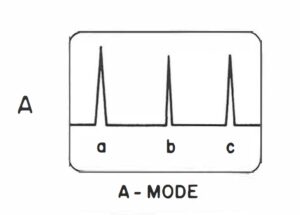
A) A – Mode ( Amplitude – Mode):- Echoes are displayed as spikes projecting from a baseline is called A – mode. The baseline identify the central axis of the beam. The spike height proportional to the echo intensity. The strong echoes producing large spikes. The probe is held stationary and pulses of nanosecond duration is sent into body and echo is generated, then the echo spikes from subsequent pulse will fall in the same position as those from the initial pulse.
In A – mode the display on the cathode ray tube contains information about the depth of structures and the amplitude of returning echo. The cathode ray tube will display about 40 dB of amplitude information corresponding to a variation in echo amplitude of about 100:1.
A – mode is used in Opthalmology, echoencephalography , echocardiography.
B) B – Mode( Brightness Mode ):- The B- mode produces a picture of a slice of tissues. B-mode is the electronic conversion of the A-mode and A-line information into brightness-modulated dots on a display screen. In general, the brightness of the dot is proportional to the echo signal amplitude. The B-mode display is used for TM-mode and 2D gray-scale imaging.
In B – Mode scanning techniques, the transducer is placed on the patient skin with acoustic coupling between tissues and transducer. The transducer may be left in one spot and rocked back and fourth, producing a simple sector scan. The transducer is moved across the patients body and rotated, producing a compound contact scan. If the angle between the transducer surface and the interface to be imaged is greater than about 5° . The amount of reflected ultrasound returning to it’s transducer will be to little to produce an image.

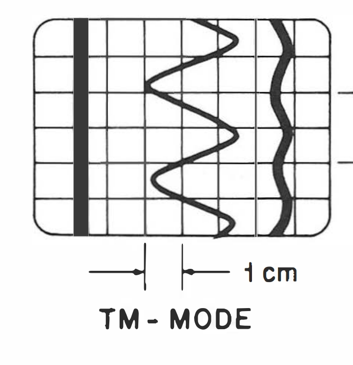
C) TM – Mode( Time Motion Mode ):- In TM – Mode , the spikes are converted in to dots. The dots are move back and fourth as indicated by the arrows. The M- mode is take permanent record. The motion must be recorded over period of time. This show the moving the line of dots to the top of the scope and then gradually dropping them to bottom. A record of the sweep is made with a camera using an exposure time longer then the sweep time because this is a time motion study. The sweep time in 3 sec. If long sweep time is used more beats are recorded and they are compacted from top to bottom.
It’s used for echocardiography and obstetrics.
FAQs
What is the principle of M mode display?
The principle of M-mode is defined as time motion display of the ultrasound wave along a chosen ultrasound line.
What is the different types of mode display?
There are 3 types of mode display in ultrasound. A- Mode , B- Mode, M- mode.