
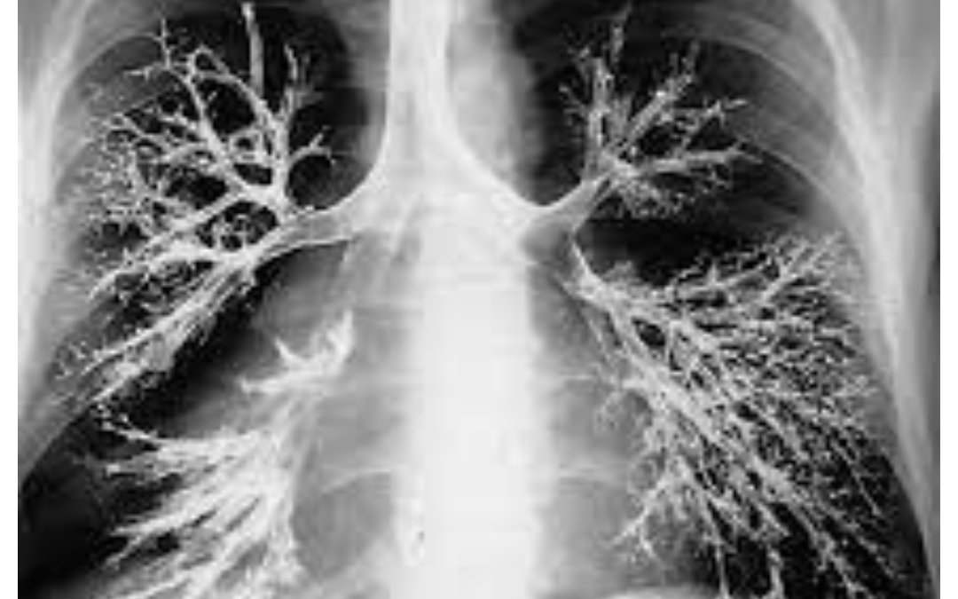
What is bronchography?
The bronchography is a radiological examination to help the study of bronchia tree and lungs by direct injection of contrast media.
There are two main types of bronchography:
A. Conventional Bronchography:- In this method, a contrast medium is introduced into the bronchial tree through a catheter that is inserted into the patient’s trachea (windpipe). The contrast medium fills the airways, and X-ray images are taken to visualize the bronchial tree and detect any abnormalities or diseases affecting the airways, such as tumors, strictures, or bronchiectasis.
B. Computed Tomography (CT) Bronchography:- This is a more advanced and non-invasive technique that uses computed tomography (CT) scanning to produce detailed cross-sectional images of the bronchial tree. Instead of a catheter, the contrast medium is usually administered through inhalation or injected intravenously. CT bronchography provides more detailed and three-dimensional images, which can be particularly helpful in diagnosing complex airway conditions.
Bronchography has been largely replaced by more advanced imaging techniques, such as CT scans and bronchoscopy, which offer better visualization and fewer risks to the patient. Bronchoscopy, for instance, allows direct visualization of the airways using a flexible scope, and it can also be used for diagnostic and therapeutic purposes. However, bronchography historically played a significant role in the diagnosis of various lung diseases and contributed to our understanding of respiratory disorders.
Indications Of Bronchography.
– Bronchiectasis.
– Bronchial obstruction
– Fistula
– Chronic bronchitis.
– Hemoptysis.
– Pulmonary lesion.
– Stenosis.
– Chronic pneumonia.
– Tumors.
– Pneumonia.
– Emphysema.
Contraindications.
– Asthma.
– High fever.
– Severe hypertension.
– weakness.
– Respiratory or cardiac disease.
– Infection.
– Poor respiratory reserve.
– Hypersensitivity to contrast media.
Contrast media.
– Use only oily contrast media.
– Dionosil ( LOCM)
Dose 20ml/ side.
Equipment.
– Fluoroscopy unit.
– Spot film device.
– Xylocaine spray.
– Xylocaine Gel.
– Bronchoscopy or catheter.
– Syringe.
Patient preparation.
– Patient must be NPO 5- 6 hrs before examination.
Preliminary Film.
– Chest PA views.
– Chest lateral view.
Procedure / technique
– When the patient come in radiology department:-
– Ask the patient to remove the all clothes and wear a hospital gown.
– The patient lies supine position on the fluoroscopy table.
– The Xylocaine spray apply for sedative the respiratory tract.
– The flexible bronchoscopy lubricant to the Xylocaine Gel.
– The flexible bronchoscopy introduce through the nose or mouth into the trachea under the observation of Fluoroscopy.
– The contrast loaded syringe attached to the catheter and 20-25 ml contrast injected into lungs.
– Then film are taken.
Filming.
A) For Right Lung.
– PA
– Right Anterior oblique (RAO).
– Right posterior oblique (RPO).
– Lateral.
B) For Left Lung.
– PA
– Left anterior oblique (LAO).
– Left posterior oblique (LPO).
– Lateral.


Aftercare.
– Patient must be advise to coughing and chest physiotherapy for removing the contrast media from chest.
– Patient must be keep in NPO till the effect of anaesthesia.
– Patient must be take an antibiotic in order to prevent infection.
Complications.
– Pain
– Bleeding.
– Pneumonia.
– Difficulty in breathing.
– Chest infection.
FAQs.
Q. What is Bronchography?
Bronchography is a medical imaging technique used to visualize the bronchial tree, the network of airways within the lungs, using a contrast medium.
Q. How is bronchography performed?
Bronchography is typically performed by introducing a contrast medium into the airways through a catheter or using CT scanning with contrast.
Q. Is bronchography safe?
Bronchography carries some risks, but complications are relatively rare. The benefits usually outweigh the risks when performed by skilled professionals.
Q. Why is bronchography performed?
Bronchography helps diagnose and assess lung conditions like tumors, bronchiectasis, and airway abnormalities.
Q. What are the types of bronchography?
The two main types of bronchography are conventional bronchography and computed tomography (CT) bronchography.
Q. How is CT bronchography different from conventional bronchography?
CT bronchography uses CT scanning to produce detailed 3D images of the bronchial tree without catheter insertion.
Q. Does bronchography require anesthesia?
In most cases, bronchography does not require general anesthesia. Local anesthesia or conscious sedation is often sufficient.
Q. How long does bronchography take?
The procedure duration varies but usually takes around 30 minutes to an hour.
Q. Is bronchography painful?
The procedure may cause mild discomfort, but it is generally not painful.
Q. What can patients expect after bronchography?
After the procedure, patients may experience mild coughing or a sore throat, but these symptoms typically resolve quickly.
Q. Can bronchography detect lung cancer?
Yes, bronchography can help identify lung tumors and assess their extent and location.
Q. Are there any risks associated with bronchography?
While rare, potential risks include allergic reactions to the contrast medium, infection, or damage to the airways.
Q. How should patients prepare for bronchography?
Patients may need to fast before the procedure and follow specific instructions provided by their healthcare provider.
Q. What are the benefits of CT bronchography over traditional bronchography?
CT bronchography provides more detailed and three-dimensional images, enabling better visualization and diagnosis.
Q. Can bronchography diagnose chronic bronchitis?
Yes, bronchography can help diagnose chronic bronchitis and assess its severity.
Q. Is bronchography suitable for children?
Bronchography is generally safe for children but is typically reserved for cases where other imaging methods are inconclusive.
Q. Are there alternatives to bronchography?
Yes, alternatives include bronchoscopy, CT scans, and magnetic resonance imaging (MRI) for assessing lung conditions.
Q. Can bronchography detect pulmonary fibrosis?
Yes, bronchography can aid in diagnosing pulmonary fibrosis and assessing its extent.
Q. How experienced should the healthcare provider be to perform bronchography?
Bronchography should be performed by experienced radiologists or interventional pulmonologists.
Q. Is bronchography covered by health insurance?
Bronchography is usually covered by health insurance, but coverage may vary, so it’s essential to check with the insurance provider beforehand.
Q. Can bronchography detect foreign bodies in the airways?
Yes, bronchography can help identify and locate foreign bodies lodged in the airways.
Q. What is the difference between bronchography and bronchoscopy?
Bronchography is an imaging technique, while bronchoscopy involves direct visualization of the airways using a flexible scope.
Q. Can bronchography diagnose asthma?
Bronchography may not be the primary method for diagnosing asthma, but it can help rule out other respiratory conditions.
Q. How much does bronchography cost?
The cost of bronchography varies depending on the healthcare facility and location.
Q. Is bronchography invasive?
Bronchography can be minimally invasive (CT bronchography) or slightly invasive (conventional bronchography with catheter insertion).
Q. Can bronchography detect lung infections?
Yes, bronchography can reveal signs of lung infections, such as pneumonia or bronchitis.
Q. Can bronchography diagnose tuberculosis?
Bronchography may contribute to diagnosing tuberculosis in certain cases but is not the primary diagnostic method.
Q. What are the contraindications for bronchography?
Contraindications may include severe respiratory distress, uncontrolled bleeding disorders, or significant allergies to contrast media.
Q. Is bronchography widely available?
Bronchography is typically available in larger medical centers or specialized imaging facilities.
Q. How soon can patients resume normal activities after bronchography?
Most patients can resume normal activities shortly after the procedure, but it’s best to follow the advice of the healthcare provider.
FOR MORE SPECIAL PROCEDURE CLICK HERE
BOOK LINK :- Fundamentals of Special Radiographic Procedures
, Excellent Site ✅✅✅