

What Is 8 Week Ultrasound?
“8 week ultrasound” also known as an early pregnancy scan or dating scan, is a medical procedure that uses ultrasound technology to visualize and assess the development of a fetus in the womb. It is typically performed around eight weeks into pregnancy, counting from the first day of the woman’s last menstrual period.
During the ultrasound, a transducer is gently moved over the woman’s abdomen or inserted into the vagina (transvaginal ultrasound) to produce sound waves. These sound waves bounce off the structures in the womb and create an image of the fetus on a monitor.
8 Week Ultrasound Provide Very Important Information Like
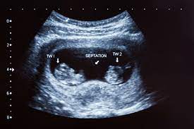
— Detection of multiple pregnancies:- At 8 weeks, it is possible to see if there are one or more embryos present, indicating if it is a single or multiple (twins/triplets) pregnancy.
— Assessment of embryonic development:- The early organ development such as the limbs, heart, brain, stomach, kidneys etc. can be observed. This confirms if the development of the embryo is as expected for 8 weeks.
— Checking for ectopic pregnancy:- The ultrasound at this stage also rules out an ectopic pregnancy by confirming that the embryo is inside the uterus. An ectopic pregnancy is a life-threatening condition and needs immediate treatment.
— Assessing potential risks:-The 8 week ultrasound also helps assess any risks or abnormalities early on based on the appearance and development of the embryo. Further tests may be recommended in some cases.
— Fetal heartbeat:- The ultrasound can detect the baby’s heartbeat, which usually becomes visible around this time.
Measurement Of 8 Week Ultrasound.
During an 8 Week ultrasound the following measurements are typically taken to assess gestational age and embryonic development:
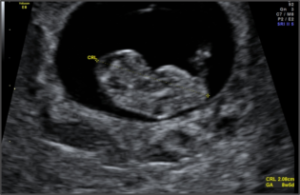
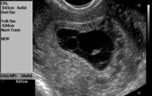
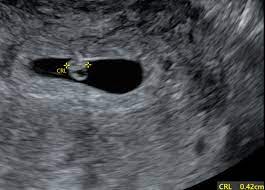
A) Crown-rump length (CRL):- This measures the length of the embryo from the top of its head (crown) to its rump (bottom). At 8 weeks, the CRL is usually around 0.63 to 0.79 inches (16 to 20 mm). This is the most accurate measurement for dating a pregnancy at this stage.
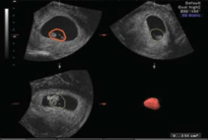
B) Gestational sac size:- The gestational sac is the fluid-filled structure that surrounds the embryo. At 8 weeks, the normal gestational sac size is usually around 0.75 to 1.5 inches (20 to 40 mm) in diameter.
C) yolk sac size:- The yolk sac provides nourishment to the embryo until the placenta forms. At 8 weeks, a normal yolk sac has a diameter of around 0.16 inches (4 mm). An enlarged or irregular yolk sac may indicate a problem with the pregnancy.
D) Fetal heart rate (FHR):- A heartbeat is usually visible at around 6-7 weeks gestation on ultrasound. At 8 weeks, a normal fetal heart rate ranges from 120 to 170 beats per minute. An abnormally slow or fast heart rate may signal certain developmental issues.

E) Arm and leg development:- At 8 weeks, small limb buds that will develop into arms and legs become visible. This confirms proper progress of embryonic development at this stage.
F) Placental and fetal pole development:- The early development of the placental tissue and fetal pole, the thickening of the embryo where limbs and head develop, is observed. Abnormal development of fetal pole and placenta may indicate growth problems.
G) Number of embryos:- In the case of multiple pregnancies, the number of embryos present is counted to confirm if it is twins, triplets or more. Each baby is measured and heart rate recorded to assess the viability and development of each embryo.
Fetal Development Of 8 Week ultrasound.
The fetal development of 8 week ultrasound are given below:-
A) Their nose is starting to show. It will look short and rather snubbed.
B) Lips.
C) The inner ear and tongue.
D) The upper jaw and roof of the mouth are coming together.
E) Their eyes are forming, but they are well protected by a layer of skin.
F) Leg and arm buds are forming.
G) Webbed fingers.
H) Their little tail is becoming smaller.
Abnormalities Of 8 Week Ultrasound.
Some common abnormalities that can be detected during 8 week ultrasound given below:-
A) Miscarriage:- If there is no visible embryo heartbeat seen, it may indicate a missed miscarriage. A follow up scan is usually done to confirm.
B) Ectopic pregnancy:- If no embryo is seen in the uterus, it may be an ectopic pregnancy located outside the uterus. This is a medical emergency.
C) Slow embryonic heart rate:- An abnormally slow heart rate below 120 beats per minute may indicate problems with the development of the embryo that could lead to possible miscarriage. A repeat scan will be done to monitor the heart rate.
D) Enlarged yolk sac:- An enlarged or irregular shaped yolk sac may indicate a problem with embryonic development and increased risk of miscarriage or fetal growth issues later on. Close monitoring with repeat ultrasounds is usually recommended.
E) Problems with limb or skull development:- Improper growth of the limb buds or skull at 8 weeks gestation may indicate a neural tube defect or genetic disorder. Further testing such as maternal blood tests and amniocentesis may be offered.
F) Improper embryonic growth:- If the crown-rump length and other measurements do not correspond to 8 weeks gestation, it may indicate improper growth of the embryo which can lead to miscarriage or growth problems later on. A repeat scan in 2 weeks is usually advised to monitor growth.
G) Multiple pregnancy complications:- In the case of twins or triplets, abnormalities such as growth problems in one embryo or a demised embryo may be detected. Close monitoring of the remaining embryos is required.
H) Placental or gestational sac problems:- Abnormal appearance or development of the placenta and gestational sac at this stage may also indicate possible problems with embryonic development or increased pregnancy loss risk. Repeat ultrasound is recommended for follow up.
I) Uncertain viability:- In some cases, it may be too early to confirm viability and a repeat scan in 1-2 weeks is needed to properly assess embryonic development, heart rate and confirm fetal viability. An uncertain viability finding at 8 weeks does not necessarily mean non-viability. But close monitoring is prudent.
J) Small crown-rump length:- Between six and eight weeks of gestation, the crown-rump length is the measurement of the entire length of the embryo. Smaller than expected fetuses at this gestational age may mean there is a concern for miscarriage.
FAQs.
Q) Can you hear a heartbeat at 8 weeks?
A stethoscope or handheld doppler devices may be used to hear the heartbeat beginning around 8 weeks.
Q) Can you see an 8 week pregnancy on ultrasound?
Yes you can see 8 weeks pregnancy on ultrasound.
Q) Can an 8 week scan detect abnormalities?
Yes, Major abnormalities of the central nervous system can be detected as early as 8 weeks of gestation.
Q) How does a 8 week fetus look?
Baby’s arms, legs, fingers and toes are all becoming more defined, and baby is less curled up, so you can see their constant little twitches and bounces. There is now an identifiable nose and upper lip, and wee little eyelids and ears.
FOR MORE CLICK HERE
BOOK LINK :- Ultrasound: The Requisites: The Requisites (Requisites in Radiology)
Very informative
Thank you
Nice presentation